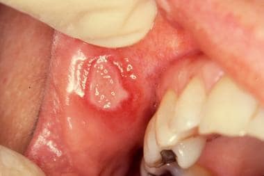
How do you treat aphthous stomatitis ?
APHTHOUS STOMATITIS :
These ulcers occur periodically and heal completely between attacks. In the majority of cases, the individual ulcers last about 7–10 days, and ulceration episodes occur 3–6 times per year. Most appear on the non-keratinizing epithelial surfaces in the mouth – i.e. anywhere except the attached gingiva, the hard palate and the dorsum of the tongue – although the more severe forms, which are less common, may also involve keratinizing epithelial surfaces. Symptoms range from a minor nuisance to interfering with eating and drinking. The severe forms may be debilitating, even causing weight loss due to malnutrition. Aphthous stomatitis, also known as a canker sore, refers to small, painful ulcers that can appear on the inside of the lips, cheeks, or soft palate; on the floor of the mouth; on the gingiva of the teeth; or on the tongue. They typically only form on non-keratinized oral mucosa, which is the cell layer that lines the inner oral cavity. This is in contrast to ulcerations from herpes simplex virus (HSV), also known as cold sores, which affect the keratinized mucosal surfaces, such as the outer surfaces of the lips. Unlike cold sores, canker sores are non-contagious.WHAT ARE THE CAUSES OF APHTHOUS STOMATITIS ?
The exact cause of aphthous stomatitis is not currently known; however, there are many factors that are thought to contribute, including a weakened immune system; emotional or psychological stress; certain foods, such as coffee, chocolate, cheese, nuts, and citrus fruits; trauma to the mouth; viral and bacterial infection; poor nutrition; and certain medications, such as aspirin, beta-blockers, chemotherapy medicines, penicillamine, sulfa drugs, and phenytoin. Sensitivity to ingredients in toothpaste and oral hygiene products, like sodium lauryl sulfate, may also trigger a canker sore. Other suspected causes include exposure to toxins (e.g., nitrites in drinking water), menstruation, or alterations in the oral microbiome. As many as 20% of cases can be related to deficiencies in iron, folate, and vitamin B6 and B12, although other deficiencies, such as vitamin D, zinc, or thiamine, may also be involved. Evidence for the T cell-mediated mechanism of mucosal destruction is strong, but the exact triggers for this process are unknown and are thought to be multiple and varied from one person to the next. This suggests that there are a number of possible triggers, each of which is capable of producing the disease in different subgroups. In other words, different subgroups appear to have different causes for the condition. These can be considered in three general groups, namely primary immuno-dysregulation, decrease of the mucosal barrier and states of heightened antigenic sensitivity (see below). Risk factors in aphthous stomatitis are also sometimes considered as either host-related or environmental.The thickness of the mucosa may be an important factor in aphthous stomatitis. Usually, ulcers form on the thinner, non-keratinizing mucosal surfaces in the mouth. Factors which decrease the thickness of the mucosa increase the frequency of occurrence, and factors which increase the thickness of the mucosa correlate with decreased ulceration.
The nutritional deficiencies associated with aphthous stomatitis (vitamin B12, folate, and iron) can all cause a decrease in the thickness of the oral mucosa (atrophy).
Local trauma is also associated with aphthous stomatitis, and it is known that trauma can decrease the mucosal barrier. Trauma could occur during injections of local anesthetic in the mouth, or otherwise during dental treatments, frictional trauma from a sharp surface in the mouth such as broken tooth, or from tooth brushing.
WHAT ARE THE SIGN AND SYMPTOMS OF APHTHOUS STOMATITIS ?
Persons with aphthous stomatitis have no detectable systemic symptoms or signs (i.e., outside the mouth). Generally, symptoms may include prodromal sensations such as burning, itching, or stinging, which may precede the appearance of any lesion by some hours; and pain, which is often out of proportion to the extent of the ulceration and is worsened by physical contact, especially with certain foods and drinks (e.g., if they are acidic or abrasive). Pain is worst in the days immediately following the initial formation of the ulcer, and then recedes as healing progresses. If there are lesions on the tongue, speaking and chewing can be uncomfortable, and ulcers on the soft palate, back of the throat, or esophagus can cause painful swallowing.Signs are limited to the lesions themselves.Ulceration episodes usually occur about 3–6 times per year. However, severe disease is characterized by virtually constant ulceration (new lesions developing before old ones have healed) and may cause debilitating chronic pain and interfere with comfortable eating. In severe cases, this prevents adequate nutrient intake leading to malnutrition and weight loss.
Aphthous ulcers typically begin as erythematous macules (reddened, flat area of mucosa) which develop into ulcers that are covered with a yellow-grey fibrinous membrane that can be scraped away. A reddish "halo" surrounds the ulcer. The size, number, location, healing time, and periodicity between episodes of ulcer formation are all dependent upon the subtype of aphthous stomatitis.
HOW IS APHTHOUS STOMATITIS IS DIAGNOSED ?
Aphthous stomatitis is usually diagnosed based on a complete history and physical examination of your child. The lesions are unique and usually allow for a diagnosis simply on physical examination. In addition, your child's doctor may order the following tests to help confirm the diagnosis and rule out other causes for the ulcers:
Blood tests
Cultures of the lesions
Biopsy of the lesion--taking a small piece of tissue from the lesion and examining it microscopically
Specific treatment for aphthous stomatitis will be determined by your child's doctor based on:
Your child's age, overall health, and medical history
Extent of the disease
Your child's tolerance for specific medications, procedures, or therapies
Expectations for the course of the disease
Your opinion or preference
The goal of treatment for aphthous stomatitis is to help decrease the severity of the symptoms. Since it is not a viral or bacterial infection, antiviral medications and antibiotics are ineffective. Treatment may include:
Increased fluid intake
Acetaminophen for any fever or pain
Proper oral hygiene
Topical medications (to help decrease the pain of the ulcers)
Mouth rinses (to help ease the pain)
It is especially important for your child to avoid spicy, salty, or acidic foods, which may cause further mouth irritation.
The vast majority of people with aphthous stomatitis have minor symptoms and do not require any specific therapy. The pain is often tolerable with simple dietary modification during an episode of ulceration such as avoiding spicy and acidic foods and beverages.Many different topical and systemic medications have been proposed (see table), sometimes showing little or no evidence of usefulness when formally investigated.Some of the results of interventions for RAS may in truth represent a placebo effect. No therapy is curative, with treatment aiming to relieve pain, promote healing and reduce the frequency of episodes of ulceration.
Medication
The first line of therapy for aphthous stomatitis are topical agents rather than systemic medication with topical corticosteroids being the mainstay treatment. Systemic treatment is usually reserved for severe disease due to the risk of adverse side effects associated with many of these agents. A systematic review found that no single systemic intervention was found to be effective. Good oral hygiene is important to prevent secondary infection of the ulcers.
Occasionally, in females where ulceration is correlated to the menstrual cycle or to birth control pills, progestogen or a change in birth control may be beneficial. Use of nicotine replacement therapy for people who have developed oral ulceration after stopping smoking has also been reported. Starting smoking again does not usually lessen the condition. Trauma can be reduced by avoiding rough or sharp foodstuffs and by brushing teeth with care. If sodium lauryl sulfate is suspected to be the cause, avoidance of products containing this chemical may be useful and prevent recurrence in some individuals. Similarly patch testing may indicate that food allergy is responsible, and the diet modified accordingly. If investigations reveal deficiency states, correction of the deficiency may result in resolution of the ulceration. For example, there is some evidence that vitamin B12 supplementation may prevent recurrence in some individuals.


Comments
Post a Comment