Should melanocytic nerus be removed ?
MELANOCYTIC NEVUS :
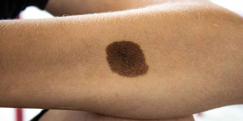
A melanocytic nevus (also known as nevocytic nevus, nevus-cell nevus and commonly as a mole is a type of melanocytic tumor that contains nevus cells. Some sources equate the term mole with "melanocytic nevus", but there are also sources that equate the term mole with any nevus form. The high concentration of the body's pigmenting agent, melanin, is responsible for their dark color . Moles are a member of the family of skin lesions known as nevi and can occur in all mammalian species, but have been documented most extensively in humans, dogs, and horses. The majority of moles appear during the first two decades of a person's life, with about one in every 100 babies being born with moles. Acquired moles are a form of benign neoplasm, while congenital moles, or congenital nevi, are considered a minor malformation or hamartoma and may be at a higher risk for melanoma. A mole can be either subdermal (under the skin) or a pigmented growth on the skin, formed mostly of a type of cell known as a melanocytic nervus .
WHAT ARE THE SIGNS AND SYMPTOMS OF MELANOCYTIC NEVUS ?
According to the most common types of moles are skin tags, raised moles and flat moles. Benign moles are usually brown, tan, pink or black (especially on dark-colored skin). They are circular or oval and are usually small (commonly between 1–3 mm), though some can be larger than the size of a typical pencil eraser (>5 mm). Some moles produce dark, coarse hair. Common mole hair removal procedures include plucking, cosmetic waxing, electrolysis, threading and cauterization.
Aging
Moles tend to appear during early childhood and during the first 30 years of life. They may change slowly, becoming raised, changing color or gradually fading. Most people have between 30 and 40 moles, but some have as many as 600.
The number of moles a person has was found to have a correlation with telomere length. However, the relation between telomeres and aging remains uncertain.
Complications
The American Academy of Dermatology says that the vast majority of moles are benign.Data on the chances of transformation from melanocytic nevus to melanoma is controversial, but it appears that about 10% of malignant melanomas have a precursor lesion, of which about 10% are melanocytic nevi. Therefore, it appears that malignant melanoma quite seldom (1% of cases) has a melanocytic nevus as a precursor.
WHAT ARE THE CAUSES OF MELANOCYTIC NEVUS ?

The cause is not clearly understood, but is thought to be caused by a defect in embryologic development. This is in the first twelve weeks of pregnancy. The defect is thought to cause a proliferation of melanocytes. This means melanocytes, the cells in the body in charge of normal skin color, are being produced at an extremely fast rate, thus causing the melanocytes to form in clusters instead of spreading out, causing abnormal skin pigmentation in some areas of the body.
Genetics
Genes can have an influence on a person's moles.
Dysplastic nevus syndrome is a largely hereditary condition which causes a person to have a large quantity of moles (often 100 or more) with some larger than normal or atypical. This often leads to a higher risk of melanoma, a serious skin cancer. Dysplastic nevi are more likely than ordinary moles to become cancerous. Dysplastic nevi are common, and many people have a few of these abnormal moles. Having more than 50 ordinary moles increases the risk of developing melanoma.
In the overall population, a slight majority of melanomas do not form in an existing mole, but rather create a new growth on the skin. Somewhat surprisingly, this also applies to those with dysplastic nevi. They are at a higher risk of melanoma occurring not only where there is an existing mole, but also where there are none. Such persons need to be checked regularly for any changes in their moles and to note any new ones.
Sunlight
Ultravoilet light from the sun causes premature aging of the skin and skin damage that can lead to melanoma. Some scientists hypothesize that overexposure to UV, including excessive sunlight, may play a role in the formation of acquired moles. However, more research is needed to determine the complex interaction between genetic makeup and overall exposure to ultraviolet light. Some strong indications that this is so (but falling short of proof), are:
- The relative lack of moles on the buttocks of people with dysplastic nevi.
- Freckles (spots of melanin on the skin, and distinct from moles) are known to be influenced by sunlight.
Studies have found that sunburns and too much time in the sun can increase the risk factors for melanoma. This is in addition to those who have dysplastic nevi being at higher risk of this cancer (the uncertainty is in regard to acquiring benign moles). To prevent and reduce the risk of melanoma caused by UV radiation, recommends staying out of the sun between 10 a.m. and 4 p.m. standard time (or whenever one's shadow is shorter than one's height). The National Cancer Institute also recommends wearing long sleeves and trousers, hats with a wide brim, sunscreens, and sunglasses that have UV-deflecting lenses.
HOW IS A MELANOCYTIC NERUS DIAGNOSED ?
Melanocytic naevi are usually diagnosed clinically by their typical appearance. If there is any doubt about the diagnosis, an expert may be consulted in person or with the help of clinical and dermatoscopic images. This is especially important if:
- A naevus changes size, shape, structure or colour
- A new naevus develops in adult life (> 40 years)
- It appears different from the person’s other naevi (a so-called ugly duckling)
- It has ABCD characteristics (Asymmetry, Border irregularity, Colour variation, Diameter > 6 mm)
- It is bleeding, crusted or itchy.
Most skin lesions with these characteristics are actually harmless when evaluated by an expert using dermatoscopy. Short-term digital dermatoscopic imaging may be used in equivocal flat lesions to check for change over time. Naevi that remain suspicious for melanoma are excised for (diagnostic ). A partial biopsy is not recommended, as it may miss an area of cancerous change.
HOW TO TREAT MELANOCYTIC NERUS ?

Most melanocytic naevi are harmless and can be safely left alone. They may be removed in the following circumstances:
- To exclude cancer
- If a naevus is a nuisance: perhaps irritated by clothing, comb or razor
- Cosmetic reasons: the mole is unsightly.
Surgical techniques include:
- Excision biopsy of a flat or suspicious melanocytic naevus
- Shave biopsy of a protruding melanocytic naevus
- Electrosurgical destruction
- Laser to lessen pigment or remove coarse hair.
Most melanocytic naevi that appear in childhood remain forever. Teenagers and young adults tend to have the greatest number of naevi. There are fewer in later life because some of them slowly fade away.
To increase the chance of spotting melanoma early, recommend:
- A self-skin examination monthly
- A patient noticing a significant change in a mole or a new lesion should show this to their doctor or
- Regular skin examinations in patients with many naevi, atypical naevi, or who have had a previous skin cancer
- Total body photography and digital dermatoscopic imaging (mole mapping) for patients at high risk of melanoma, especially if they have many melanocytic naevi.
The number of melanocytic naevi can be minimised by strict protection from the sun, starting from birth. Sunscreen alone is not sufficient to prevent new naevi from appearing.
At any age, sun protection is important to reduce skin aging and the risk of skin cancer.
- In New Zealand, the SunSmart Sun Protection Alert advises when protection is required.
- Coverup . Wear a hat, long sleeves and a long skirt or trousers. Choose fabrics designed for the sun (UPF 40+) when outdoors.
- Apply sunscreen to areas you can't cover. Choose broad-spectrum high protection (SPF 50+) sunscreens, applied frequently to exposed areas.


Comments
Post a Comment