How aggressive is acral lentigious melanoma ?
ACRAL LENTIGINOUS MELANOMA :

Acral lentiginous melanoma (ALM) is a type of malignant melanoma. Malignant melanoma is a form of skin cancer that happens when the skin cells called melanocytes become cancerous. Melanocytes contain your skin color (known as melanin or pigment). In this type of melanoma, the word “acral” refers to the occurrence of the melanoma on the palms or soles.
ALM is the most common type of melanoma in people with darker skin and those of Asian descent. However, it can be seen in all skin types. ALM may be hard to recognize at first, when the patch of darkened skin is small and looks like little more than a stain or bruise. Early diagnosis and treatment are essential.
What are the sign and symptoms of Acral lentiginous melanoma ?
There are five signs that you can look for to decide whether a spot may be suspicious for melanoma (as opposed to a non-cancerous mole). These steps are easy to remember by the acronym ABCDE :
- Asymmetry: The two halves of the spot are not the same as each other, meaning that they may vary in size or shape. Non-cancerous moles are usually round in shape or are the same size and shape on both sides.
- Border irregularity: The border around the spot is uneven or jagged. Non-cancerous moles usually have borders that are straight, clearly defined, and solid.
- Color variation: The spot is made up of areas of multiple colors of brown, blue, black, or other similar colors. Non-cancerous moles are normally just one color (usually brown).
- Large diameter: The spot is larger than a quarter of an inch (0.25 inch, or 6 millimeters) around. Non-cancerous moles are typically much smaller.
- Evolving: The spot has gotten bigger or has more colors than when it originally appeared on your skin. Non-cancerous moles usually don’t grow or change color as drastically as a spot of melanoma.
The surface of a spot of ALM may also start out smooth and become bumpier or rougher as it evolves. If a tumor begins to grow from the cancerous skin cells, the skin will become more bulbous, discolored, and rough to the touch.

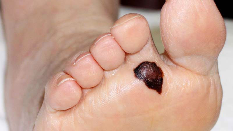
ALM can also appear around your fingernails and toenails. When this happens, it’s called subungal melanoma. You may notice general discoloration in your nail as well as spots or lines of discoloration extending onto the cuticle and skin where it meets the nail. This is called Hutchinson’s sign. As the spot of ALM grows, your nail might begin to crack or break altogether, especially as it advances to later stages. The most visible symptom of ALM is typically a dark spot of skin that’s surrounded by skin that remains your normal skin color. There’s a clear border between the dark skin and the lighter skin around it. You’ll usually find a spot like this on or around your hands and feet, or in the nail beds.
ALM spots may not always be dark-colored or even dark at all. Some spots may be reddish or orange in color — these are called amelanotic (or non-pigmented).
What are the causes of Acral lentiginous melanoma ?
ALM happens because the melanocytes in your skin become malignant. A tumor will continue to grow and spread until it’s removed.
Unlike other forms of melanoma, acral lentiginous melanoma is not associated with excess sun exposure. It’s believed that genetic mutations contribute to the development of acral lentiginous melanoma.
Acral lentiginous melanoma treatment | Treatment and management
Early stages
If your ALM is still in the early stages and is small enough, your doctor may simply be able to cut the spot of ALM out from your skin in a quick, outpatient surgical procedure. Your doctor will also cut out some skin around the area. How much skin needs to be removed depends on the Breslow thickness of the melanoma, which measures how deeply the melanoma invades. This is determined microscopically.
Advanced stages
If your ALM has a deeper level of invasion, lymph nodes may need to be removed. Amputation of digits may even be necessary. If there is evidence of distant spread, such as to other organs, you may need treatment with immunotherapy. Immunotherapy with biologic medications targets receptors in the tumor.
How is a Acral lentigious melanoma diagnosis ?
Although the ideal method of diagnosis of melanoma is complete excisional biopsy, alternatives may be required according to the location of the melanoma. Dermatoscopy of acral pigmented lesions is very difficult but can be accomplished with diligent focus. Initial confirmation of the suspicion can be done with a small wedge biopsy or small punch biopsy.Thin deep wedge biopsies can heal very well on acral skin, and small punch biopsies can give enough clue to the malignant nature of the lesion. Once this confirmatory biopsy is done, a second complete excisional skin biopsy can be performed with a narrow surgical margin (1 mm). This second biopsy will determine the depth and invasiveness of the melanoma and will help to define what the final treatment will be. If the melanoma involves the nail fold and the nail bed, complete excision of the nail unit might be required. Final treatment might require wider excision (margins of 0.5 cm or more), digital amputation, lymphangiogram with lymph node dissection, or chemotherapy


Comments
Post a Comment