How is paraneoplastic pemphigus diagnosed ?
PARANEOPLASTIC PEMPHIGUS :

Paraneoplastic pemphigus (PNP) is an autoimmune disorder stemming from an underlying tumor . It is hypothesized that antigens associated with the tumor trigger an immune response resulting in blistering of the skin and mucous membranes .
While patients with malignant and benign tumors are both at risk, malignancy is associated with high mortality rates (near 90%). Current treatment focuses on general wound healing and administering corticosteroids, which has not demonstrated a high success rate. Recent research developments aim to treat the underlying tumor in order to alleviate the symptoms of PNP.
WHAT ARE THE SIGNS AND SYMPTOMS OF PARANEOPLASTIC PEMPHIGUS ?

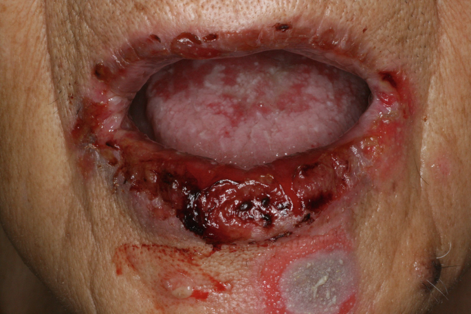
It is most common that muscous membrane lesions of the oral cavity are presented first. They can involve the oropharynx , nasopharynx tongue, and vermilion (red portion) of the lips. They are also known to develop in the conjunctive of the eye, anogenital region, and esophagua. Cutaneous lesions tend to follow the onset of mucosal lesions. The blisters often erupt in waves, usually affecting the upper trunk, head, neck, and proximal extremities. Pemphigoid-like lesions are seen more often on the extremities. Lichenoid lesions are more common among children, presenting on the trunk and limbs, ranging from small red scaly papules to extensive violet to brown papules extending to the face and neck. Within the spectrum of lichenoid presentations are wounds that have features of erythema multiforme and graft-vs.-host disease. Scaly lesions on the palms of the hand and soles of the feet have been noted to coincide with the lichenoid lesions. Lesions of varying morphology may present simultaneously and transform from one type to another as the disease progresses.
HOW IS A PARANEOPLASTIC PEMPHIGUS DIAGNOSIS ?
In order to diagnose paraneoplastic pemphigus, several tests may be performed. Initially, samples are obtained via skin biopsy for routine microscopy and direct immunofluorescence (DIF) testing. The skin sample needs to be obtained from an unaffected area adjacent to a lesion. Testing in more detail follows depending on the results from the DIF. Prompt diagnosis of PNP is crucial due to the high mortality rate of the disease.
Camisa and Helm revised the original criteria from Anhalt et al. into major and minor signs indicating PNP: Major:
- Polymorphic mucocutaneous eruption
- Concurrent internal tumor
- Serum antibodies with a specific immunoprecipitation pattern
Minor:
- Histologic evidence of acantholysis (loss of intercellular connections leading to breaking apart of the skin; lesion)
- Direct immunofluorescence showing intercellular and basement membrane staining
- Indirect immunofluorescence staining with rat bladder epithelium
Microscopy
Microscopy of the skin sample obtained from the biopsy is used to detect the presence of cleavage within the dermis, epidermal acantholysis (breaking apart of the skin), dyskeratotic keratinocytes and vacuolar changes in the layers of the skin, interfacial dermatitis and epidermal exocytosis. Presentation of these characteristics suggests PNP.
Direct immunofluorescence testing
Detection in other locations such as intercellular and areas below the epidermis (subepidermal), as well as along the dermoepidermal junction (area that joins the epidermis and dermis), suggests paraneoplastic pemphigus.
Indirect immunofluorescence (IDIF)
Patients with high concentration of antibodies show intercellular, intraepidermal antibodies as well as along the dermoepidermal junction. Patients with low concentration of antibodies only present with them inside the cells (intercellular).
If the results are negative, perform the additional assays regardless. Cases have been confirmed that reported with initial negative DIF and IDIF tests.
Similar diseases with overlapping symptoms
PNP is most commonly mistaken for pemphigus vulgaris due to the extreme similarities of the lesions that develop. However, the difference lies in the specificity of the autoreactive antibodies in each case.
RISK FACTOR OF PARANEOPLASTIC PEMPHIGUS :
As PNP is ultimately caused by the presence of a tumor, it is not contagious. There is no known way to predict who will become afflicted with it. Patients with cancer are therefore a group at risk. Although PNP has been known to affect all age groups, it is more likely to afflict middle-aged to older patients.
HOW TO TREAT PARANEOPLASTIC PEMPHIGUS ?
Wound healing
Initial treatment involves addressing any existing infections that may have occurred due to the broken state of the skin. Existing wounds are treated with warm compresses, non-adherent (non-stick) dressing, and topical antibiotic ointment. Immunosuppressive agents are administered in attempt to decrease blistering; this is not often effective. The first medication given aiming to heal the wounds are high dosecorticosteroids. This is followed by steriod sparing agents which may reduce steroid intake and therefore lessen the side effects. Skin lesions are more likely to respond to this line of treatment than mucosal lesions. However, a high level of caution is advised in patients with a confirmed malignancy, where immunosuppression is vital and dictates treatment options. If the initial therapy fails to control the symptoms of PNP, and the condition of the patient deteriorates, a more aggressive approach may be necessary.
Medication
Prednisone
Prednisone is an immunosuppressive agent which affects all of the organ systems. Effects on the cellular level include cell activation, replication, differentiation, and mobility. The overall goal is to decrease blistering (inhibition of immediate and delayed hypersensitivity) through decreasing the production of autoantibodies. In order to suppress the production of antibodies, higher doses must be administered. Lesser doses can be prescribed in order to achieve suppression of monocyst function.
Azathioprine
Azathioprine is a steroid-sparing agent used in combination with Prednisone. It functions by inhibiting RNA and DNA synthesis.
Ciclosporin
Ciclosporin is an immunosuppressive agent most often used in organ transplantation that has demonstrated to be effective with skin disorders. It functions by lessening production of autoantibodies and therefore diminishing the development of blisters and erosions. The mechanism of action is by inhibiting the production of T lymphocytes and lymphokines.
Cyclophosphamide
Cyclophosphamide is an immunomodulator used in combination with systemic steroids to remove bone marrow. This is followed by transplanting peripheral blood stem cells.


Comments
Post a Comment